Publications
Search
Type
Methods
Topics
2026
-
-
Beyond Accuracy: A Leakage-Safe and Anatomy-Aware Evaluation of Gastrointestinal Endoscopy Classification preprintartificial-intelligencedeep-learningrepresentation learning
Submitted for publication at- 29th International Conference on Computer and Information Technology. Submission ID:#2814. (2026)
ABSTRACT
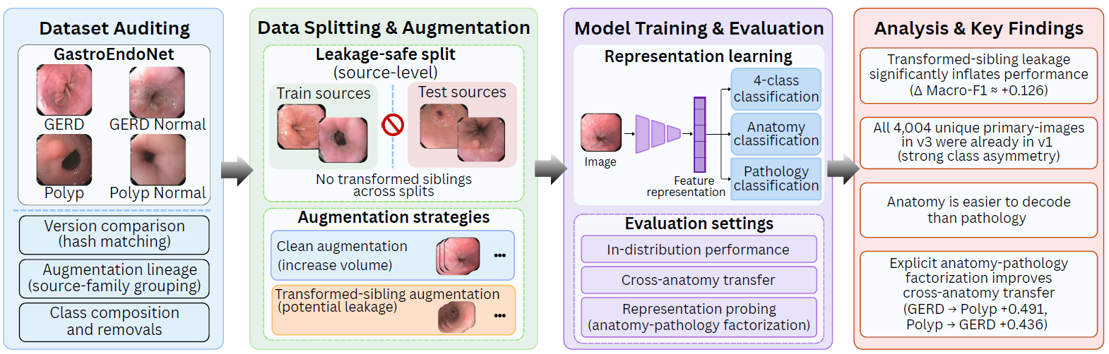
Accurate brain tumor diagnosis depends on early detection, yet most publicly available magnetic resonance imaging (MRI) datasets and the deep learning models benchmarked on them are drawn from Western populations, leaving South Asian clinical settings comparatively underserved. This paper reports a systematic benchmarking study on Publicly Available MRI Dataset for Radiological Analysis and Modeling (PMRAM), a Bangladeshi brain MRI dataset, for the four-class problem of separating glioma, meningioma, pituitary tumor and tumor-free scans. We fine-tuned seven pretrained convolutional neural networks (CNN) (ResNet18, ResNet50, VGG16, VGG19, EfficientNet-B1, MobileNetV2, and DenseNet121), each under three optimizers (AdamW, Adam, and SGD) with a cosine-annealed learning rate, giving 21 CNN configurations. To probe whether global self-attention adds value on this data, we evaluated four Vision Transformer (ViT) variants (ViT-Base, ViT-Small, DeiT-Base and BEiT-Base) under the same optimizer protocol, adding 12 further configurations. The five strongest configurations were then re-assessed with Stratified 5-Fold Cross-Validation to confirm that their accuracy was not an artifact of a single fortunate split. ResNet50 with AdamW gave the best cross-validated result, 98.74% mean accuracy, narrowly ahead of the leading transformer. Taken together, the experiments show that transfer learning, paired with disciplined validation, already reaches clinically encouraging accuracy on a modest regional dataset, and they set the stage for our planned work on explainable AI (XAI) and hybrid CNN–Transformer models.High classification accuracy in gastrointestinal (GI) endoscopy does not necessarily indicate that a model has learned transferable pathology cues, particularly when augmentation lineage, dataset provenance, and anatomical context are not explicitly controlled. This study audits GastroEndoNet, a fourclass endoscopy dataset containing gastroesophageal reflux disease (GERD), GERD Normal, Polyp, and Polyp Normal images, from dataset construction through representation learning. A sourcefamily- controlled benchmark was first used to separate the effect of clean augmentation volume from transformed-sibling leakage. Exposing training to augmented siblings of held-out sources increased Macro-F1 by approximately 0.126, whereas an equalsized clean augmentation expansion improved Macro-F1 by only about0.010. Cross-version hash analysis further showed that all 4,004 unique primary images in version3 were already present in version 1, with strongly class-asymmetric removals. Under leakage-safe evaluation, anatomy was substantially easier to decode than pathology, and disease representations transferred poorly across anatomical branches. Explicit anatomy– pathology factorization substantially improved cross-anatomy transfer, yielding GERD→Polyp and Polyp→GERD Macro-F1 gains of 0.491±0.022 and 0.436±0.021, respectively, while direct four-class Macro-F1 decreased modestly by0.018 on average. These findings show that reliable endoscopy benchmarking requires auditing source dependence, dataset provenance, and representation transferability in addition to reporting aggregate classification accuracy.
-
-
Benchmarking CNN and Vision Transformer Architectures for Four-Class Brain Tumor Classification on the PMRAM Bangladeshi MRI Dataset preprintartificial-intelligencedeep-learningclassification
Accepted for publication at-2026 IEEE International Conference on Optics, Machine Learning and Emerging Technology. (2026)
ABSTRACT
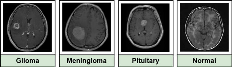
Accurate brain tumor diagnosis depends on early detection, yet most publicly available magnetic resonance imaging (MRI) datasets and the deep learning models benchmarked on them are drawn from Western populations, leaving South Asian clinical settings comparatively underserved. This paper reports a systematic benchmarking study on Publicly Available MRI Dataset for Radiological Analysis and Modeling (PMRAM), a Bangladeshi brain MRI dataset, for the four-class problem of separating glioma, meningioma, pituitary tumor and tumor-free scans. We fine-tuned seven pretrained convolutional neural networks (CNN) (ResNet18, ResNet50, VGG16, VGG19, EfficientNet-B1, MobileNetV2, and DenseNet121), each under three optimizers (AdamW, Adam, and SGD) with a cosine-annealed learning rate, giving 21 CNN configurations. To probe whether global self-attention adds value on this data, we evaluated four Vision Transformer (ViT) variants (ViT-Base, ViT-Small, DeiT-Base and BEiT-Base) under the same optimizer protocol, adding 12 further configurations. The five strongest configurations were then re-assessed with Stratified 5-Fold Cross-Validation to confirm that their accuracy was not an artifact of a single fortunate split. ResNet50 with AdamW gave the best cross-validated result, 98.74% mean accuracy, narrowly ahead of the leading transformer. Taken together, the experiments show that transfer learning, paired with disciplined validation, already reaches clinically encouraging accuracy on a modest regional dataset, and they set the stage for our planned work on explainable AI (XAI) and hybrid CNN–Transformer models.
-
-
A Dual-Pipeline Approach for Colorectal Polyp Detection and Segmentation with Machine and Deep Learning Models preprintartificial-intelligencedeep-learningsegmentation
Submitted for publication at International Journal of Intelligent Systems. Manuscript ID:7426684 (2026)
ABSTRACT
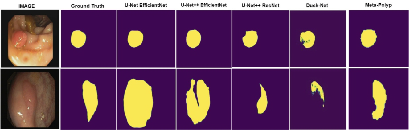
The rate of colorectal cancer is growing at an alarming rate all over the world. The survival rate of patients drastically 10decreases when the cancer reaches its final stage. Hence the identification and removal of colon polyps is very important for the patients 11 at early stages. Advanced computer-aided systems can assist polyp detection that outperforms manual detection methods. In this paper 12we present the development of an automated polyp detection system using advanced machine learning and deep learning models. The 13 first pipeline of this work involved classification of polyps using machine learning models and deep learning models. We explored two 14methods of image feature extraction for the machine learning classifiers namely, Local Binary pattern (LBP) and Gray Level Co- 15 occurrence matrix (GLCM). The LBP based feature extraction, with LightGBM classifier gave the best accuracy for polyp classification, 16 achieving 94% accuracy and 93.9% F1-score. In comparison to the machine learning classifier, the deep learning classifier EfficientNetB0 17 achieved up to 96% test accuracy. The second pipeline employed deep learning models for polyp segmentation, with the best model (U- 18 Net with the EfficientNetB0 backbone) achieving up to 90.76% and 72.13% dice coefficients on the validation and test datasets 19 respectively. Overall, we confirmed that the deep learning pipeline gave promising results for both polyp segmentation and classification.
-
-
Enhanced vascular perfusion mapping and heart rate estimation via spatio-temporal rPPG with optical and motion compensation techniques journalphysiological-monitoringsignal-processingimage-processingoptical-measurement
Biomedical Physics and Engineering Express (2026)
ABSTRACT BIB
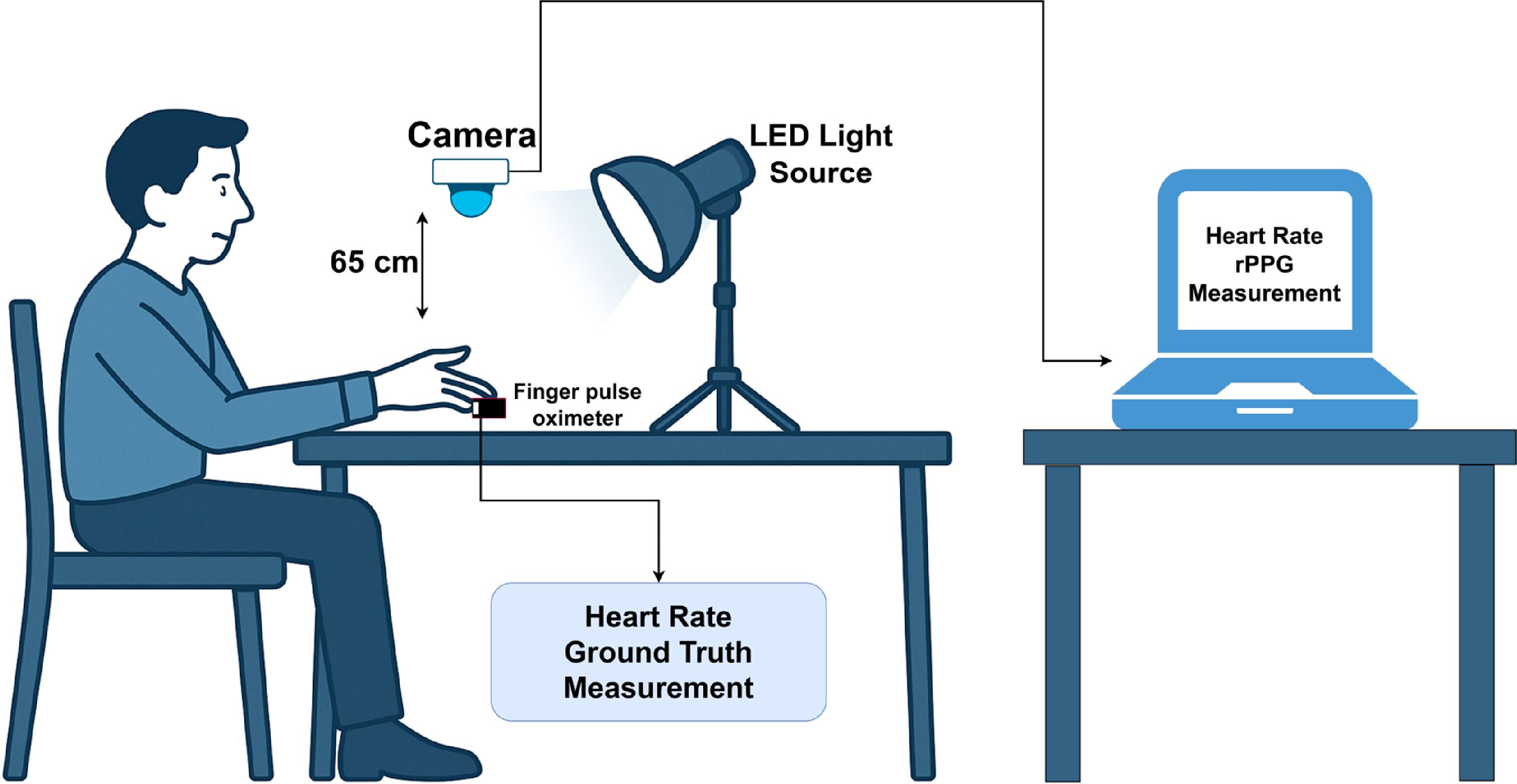
Remote photoplethysmography (rPPG) offers a non-contact method for monitoring physiological signals using camera-based systems. The goal of this research is to estimate heart rate and spatial distributions of vascular perfusion using spatio-temporal rPPG (ST-rPPG) and to evaluate the impact of polarization, spectral filtering, and motion compensation on perfusion map quality and heart rate estimation. Two acquisition setups were used: an RGB camera with and without cross-polarization, and a monochrome camera combined with spectral filters. A motion compensation strategy was implemented that combined optical flow-based stable segment selection and temporal video stabilization to reduce motion artifacts. Four rPPG algorithms (GREEN, CHROM, POS, and G-R) were evaluated using three performance metrics: Absolute Error (AE), Signal Quality Index (SQI), and Signal-to-Noise Ratio (SNR) under cross polarized and non polarized lighting in 20 subjects to assess their suitability for perfusion mapping. GREEN and G-R method stood out giving the best results. In the second setup, nine spectral filters were tested across three anatomical regions using the GREEN method, to investigate the influence of wavelength selection on spatial perfusion signal quality. Green, orange, and blue wavelengths produced the best results in terms of AE, SQI and SNR, particularly in the palm region. Visualizations like the spatial perfusion maps, confirmed the superiority of motion-compensated, polarized, and spectrally optimized conditions for enhancing non-contact vascular perfusion assessment. Prior rPPG studies focused primarily on facial datasets or single optical factors, while this work provides the systematic evaluation of polarization, spectral filtering, and motion compensation in a unified hand-based framework, extending established rPPG methods toward high-resolution perfusion mapping.
2025
-
-
Revival of Muslin by Phuti Karpas plant identification with convolution neural network journalcomputer-vision-applicationsdeep-learningimage-processing
Array (2025)
ABSTRACT BIB
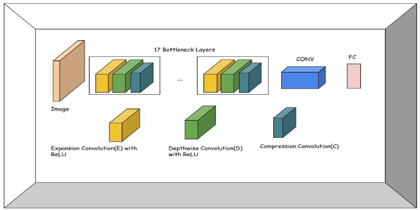
Phuti Karpas, historically central to cotton production and thought extinct, has re-emerged in botanical research, prompting a need for reliable identification methods. This study develops a systematic approach for classifying Phuti Karpas leaves using various convolutional neural networks (CNNs), including AlexNet, Inception, VGG16, MobileNetV2, and a custom-designed baseline model. A unique dataset of 2354 leaf images was curated, with two main classes: Phuti Karpas and Non Phuti Karpas, the latter including 14 other plant types to enhance model robustness. Each model was evaluated on metrics like accuracy, precision, recall, computational time, and memory efficiency. AlexNet yielded the highest average accuracy, while the custom baseline model, optimized for mobile deployment, provided comparable accuracy with faster inference. To demonstrate real-world usability, an Android app was created for real-time Phuti Karpas identification, offering an accessible tool for field researchers and conservationists. This work not only advances deep learning applications in plant taxonomy but also aids in the cultural and scientific revival of Phuti Karpas.
2023
-
-
Effect of Input Channel Reduction on EEG Seizure Detection journalbiomedical-signal-analysiscomputer-aided-diagnosismachine-learningdeep-learningsignal-processingfeature-selection
Przegląd Elektrotechniczny (2023)
ABSTRACT BIB
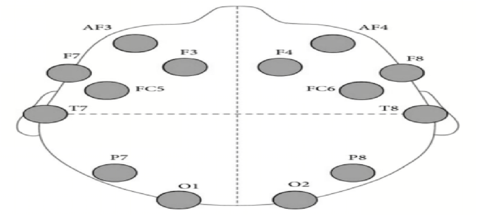
In this study, the effectiveness of six machine learning and eight deep learning algorithms in analyzing electroencephalogram (EEG) signals for detecting epileptic seizures has been investigated. The study utilizes 14 channels in the EMOTIV EPOC+ device which is based on international 10-20 system. To find out the most informative and sensitive channel, one of the 14 channels has been dropped one at a time. The accuracy values were determined for all the methods using two different datasets: the Guinea-Bissau dataset and the Nigeria dataset, both available on https://zenodo.org/record/1252141#.ZDdyj3ZByUl. In case of machine learning models, the performance of SVM classifier performs best with maximum accuracy of 83.2% (Guinea-Bissau) and 77% (Nigeria) without excluding any channels. No significant performance degradation has been observed for single channel exclusion of this classifier. Among the deep learning models, the four best performing models in terms of accuracy are CNN-LSTM (92.5%), IC-RNN (91.8%), ChronoNet (91.1%) and C-DRNN (88.6%). After excluding one channel at a time and investigating their effect on the performance of the four DL models, it has been observed that the most significant and most sensitive channels lie within the frontal and parietal zone. This finding will be very useful in practice as it indicates that the electrodes in the frontal and parietal zone should be placed with absolute precision for accurate diagnosis of the diseases. In addition, this study also explore the effectiveness of the selected classifiers in detecting seizure in case of failure of any particular EEG signal channel.
2022
-
-
ANN based approach to predict Criminal Trends in Bangladesh. conferencemachine-learning
2nd International Conference on Artificial Intelligence and Signal Processing (AISP), IEEE (2022)
ABSTRACT BIB
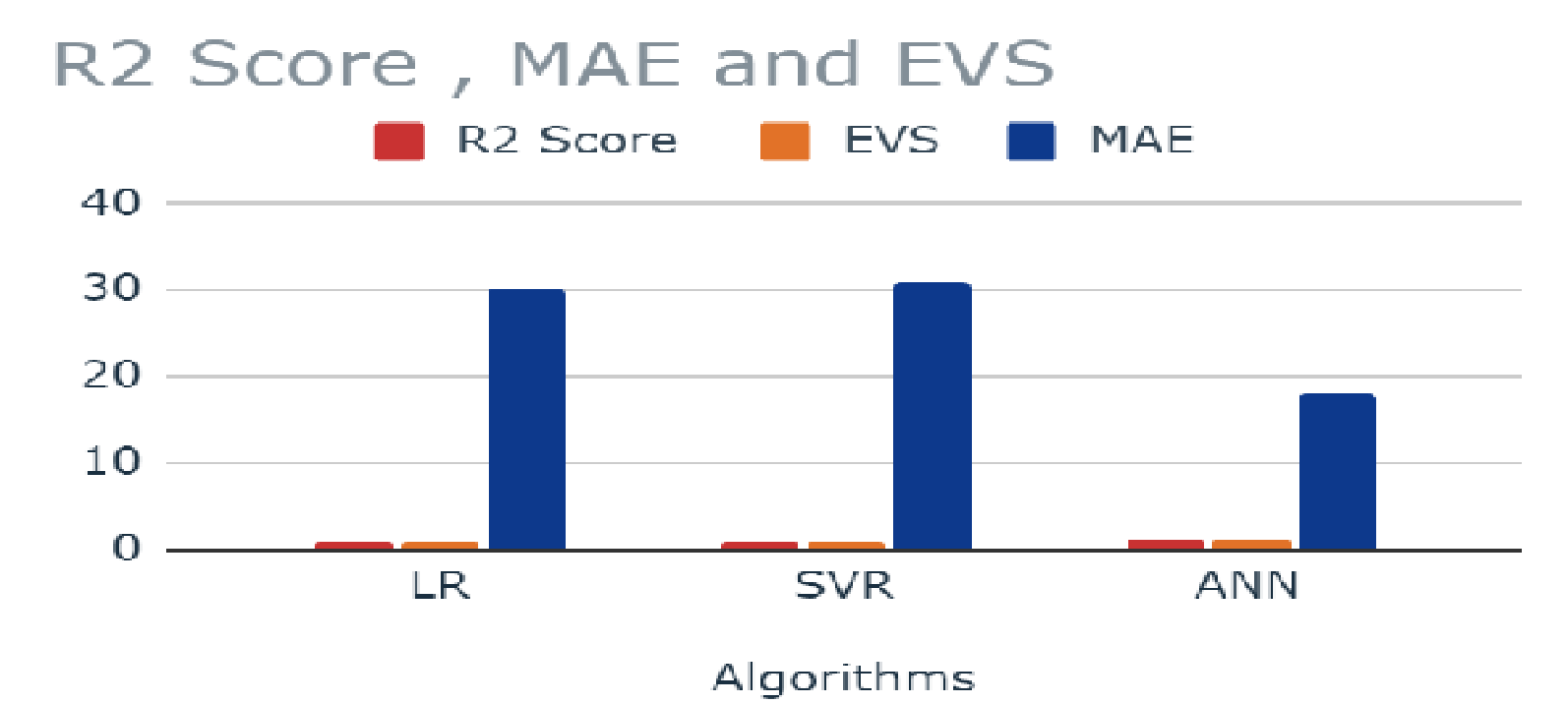
Crime trend analysis has become a mandatory task as the scale of crime is increasing rapidly all over the globe. In recent years, Bangladesh has encountered various types of crimes and the rate is increasing with its increased population. Both physical and digital based crimes have become very common and their fatality can disrupt the advancement of any country. To tackle the situation, it is important to investigate and forecast the crime patterns that can assist the law enforcement agencies for easier investigation. Machine Learning and Deep Learning based crime analysis has become very popular as they can accurately and efficiently analyze large criminal dataset. In this study, the authors implemented machine learning techniques as well as ANN architecture to assess and forecast crime trends in Bangladesh. The dataset in this experiment was collected from the Bangladesh Police website that consists of criminal records of various crimes during the years 2010 to 2019. Authors evaluated the performance of ANN with other regression models named linear Regression and Support Vector Regression for crime prediction. The proposed model(ANN) outperforms other two models in this study compared to all the performance evaluation metrics. Hence ANN model is suggested by the authors to forecast and analyze future crime trends.
-
-
High Accuracy Swin Transformers for Image-based Wafer Map Defect Detection journalcomputer-vision-applicationsdeep-learningimage-processing
International Journal of Engineering and Manufacturing (IJEM) (2022)
ABSTRACT BIB
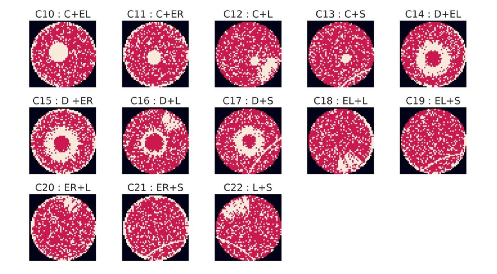
A wafer map depicts the location of each die on the wafer and indicates whether it is a Product, Secondary Silicon, or Reject. Detecting defects in Wafer Maps is crucial in order to ensure the integrity of the chips processed in the wafer, as any defect can cause anomalies thus decreasing the overall yield. With the current advances in anomaly detection using various Computer Vision Techniques, Transformer Architecture based Vision models are a prime candidate for identifying wafer defects. In this paper, the performance of Four such Transformer based models – BEiT (BERT Pre-Training of Image Transformers), FNet (Fourier Network), ViT (Vision Transformer) and Swin Transformer (Shifted Window based Transformer) in wafer map defect classification are discussed. Each of these models were individually trained, tested and evaluated with the “MixedWM38” dataset obtained from the online platform, Kaggle. During evaluation, it has been observed that the overall accuracy of the Swin Transformer Network algorithm is the highest, at 97.47%, followed closely by Vision Transformer at 96.77%. The average Recall of Swin Transformer is also 97.54%, which indicates an extremely low encounter of false negatives (24600 ppm) in contrast to true positives, making it less likely to expose defective products in the market.
2021
-
-
A Performance Evaluation of Machine Learning Models on Human Activity Identification (HAI) conferencemachine-learningdeep-learningsignal-processing
Proceedings of the 12th International Conference on Soft Computing and Pattern Recognition (SoCPaR 2020) (2021)
ABSTRACT BIB
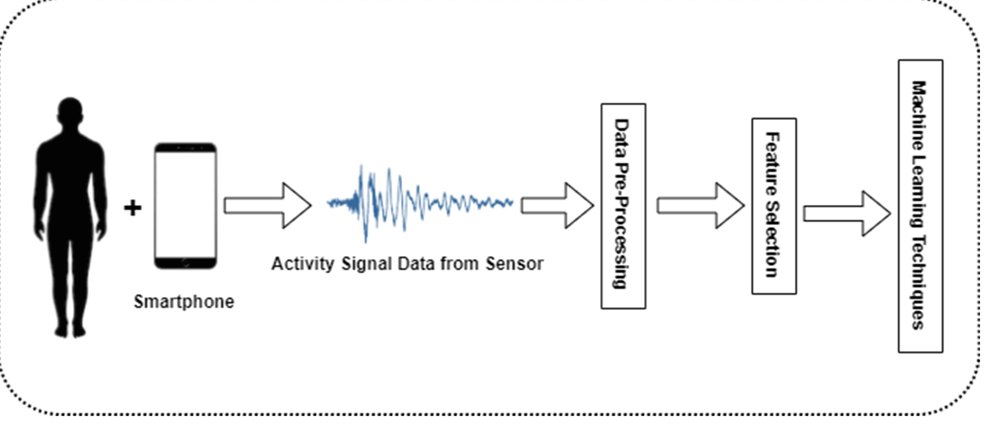
Human activity identification (HAI) is presently a promising field for artificial intelligence researchers. HAI can be applied in various areas in our daily life such as surveillance, health care, etc. There are several machine learning and deep learning techniques that are applied to recognize the human activity. Various multi-dimensional sensors from smartphones, smart tablets, smartwatches can record different types of human activity. Some extensive sensors in smartphones such as accelerometer, gyroscope, microphone, GPS, and camera which can respond to record human gestures. These gestures can be sitting, lying, standing, walking, etc. In this study, the authors analyze the performance of traditional machine learning techniques as well as 1D-CNN architecture to recognize human activity using the UCI human activity identification dataset. PCA has been enforced to reduce the dimension and feature selection to get better results. 1D-CNN performs better in this study with an accuracy of 97.30%. Where Logistic regression achieves 96.00%, Linear Support Vector Machine achieves 93.71%, Kernel-Support Vector Machine achieves 94.85%, Random Forest achieves 90.59% and Decision Tree achieves 84.46% accuracy. All the accuracies are considered by taking the average of individual class accuracy. However, the 1D-CNN model is proposed by the authors to implement in less computational powered devices such as smartphones, smartwatches, etc.
-
-
Recent Advances in Computer-Aided Medical Diagnosis Using Machine Learning Algorithms with Optimization Techniques journalcomputer-aided-diagnosismachine-learningoptimizationfeature-selection
IEEE Access (2021)
ABSTRACT BIB
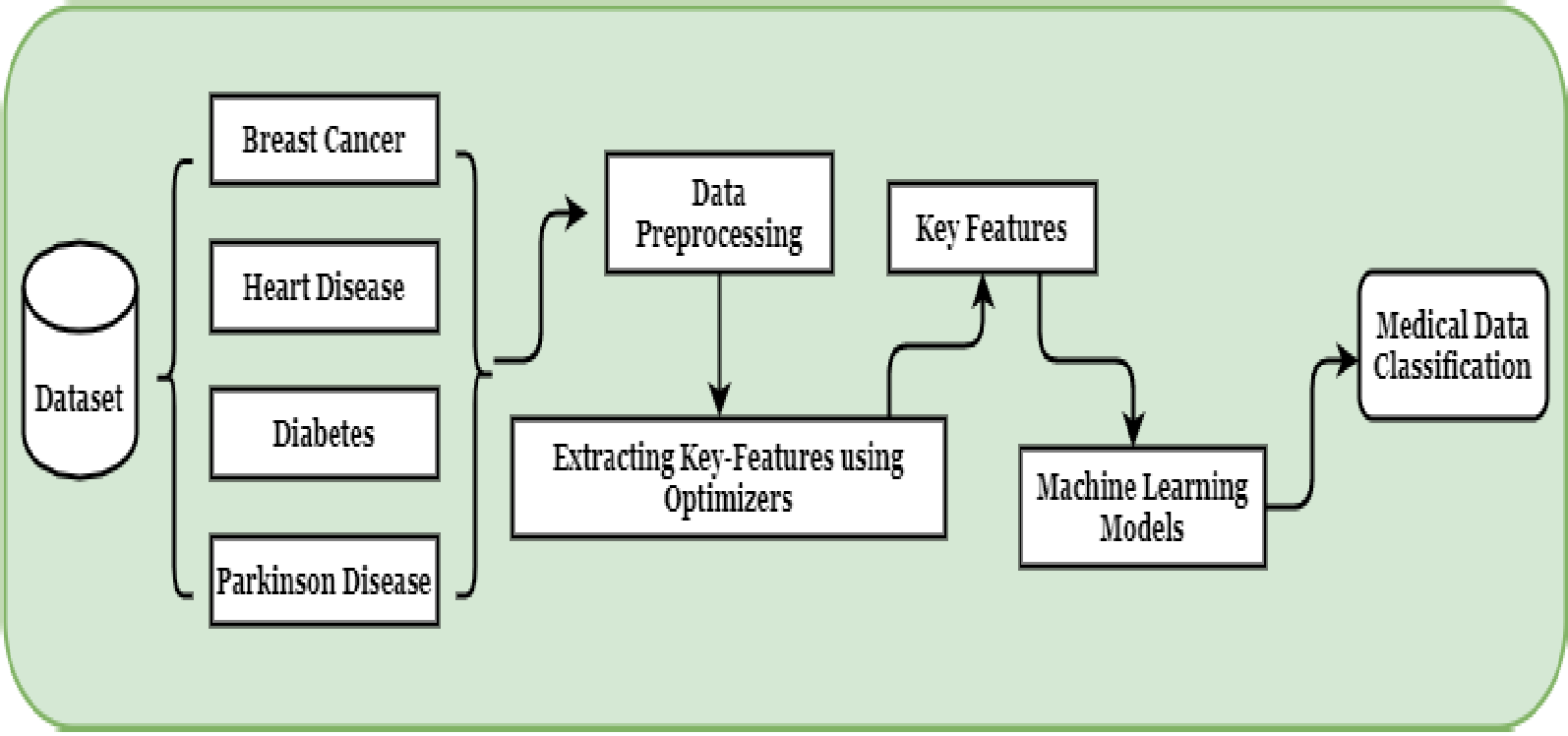
Artificial intelligence is a spectacular part of computer engineering that has earned a compelling diversion in the field of medical data classification due to its state-of-art algorithmic strength and learning capabilities. Machine Learning is a major sub-domain of artificial intelligence, where it has become one of the most promising fields in computer science. In recent years, there is a large spectrum of healthcare and biomedical data that has been growing intensely. Due to the huge labeled or unlabeled data, it is important to have a compact and robust machine learning solution for classification. Several optimizers have been deployed to improve the inclusive performance of machine learning models. The classification of machine learning models depends on several factors. This comprehensive review paper aims to insight into the current stage of optimized machine learning success on medical data classification. An increasing number of unstructured medical data has been utilizing in machine learning algorithms to predict intuitions. But it is difficult to inherent immense intuition from those data. So machine learning researchers have utilized state-of-art optimizers and novel feature selection techniques to overcome and emend the performance accuracy. We have highlighted some recent literature, which exhibits the robust impact of optimizers and feature selection on machine learning techniques on medical data characterization. On the other hand, a clean-cut introduction on machine learning and theoretical outlook of widely utilized optimization techniques like genetic algorithm, gray wolf optimization, and particle swarm optimization are discussed for initial understanding of the optimization techniques.
2020
-
-
Electroencephalogram (EEG) brainwave signal-based emotion recognition using extreme gradient boosting algorithm. journalbiomedical-signal-analysismachine-learningdeep-learningsignal-processing
Annals of Engineering, Imperial Open Publishing (2020)
ABSTRACT BIB
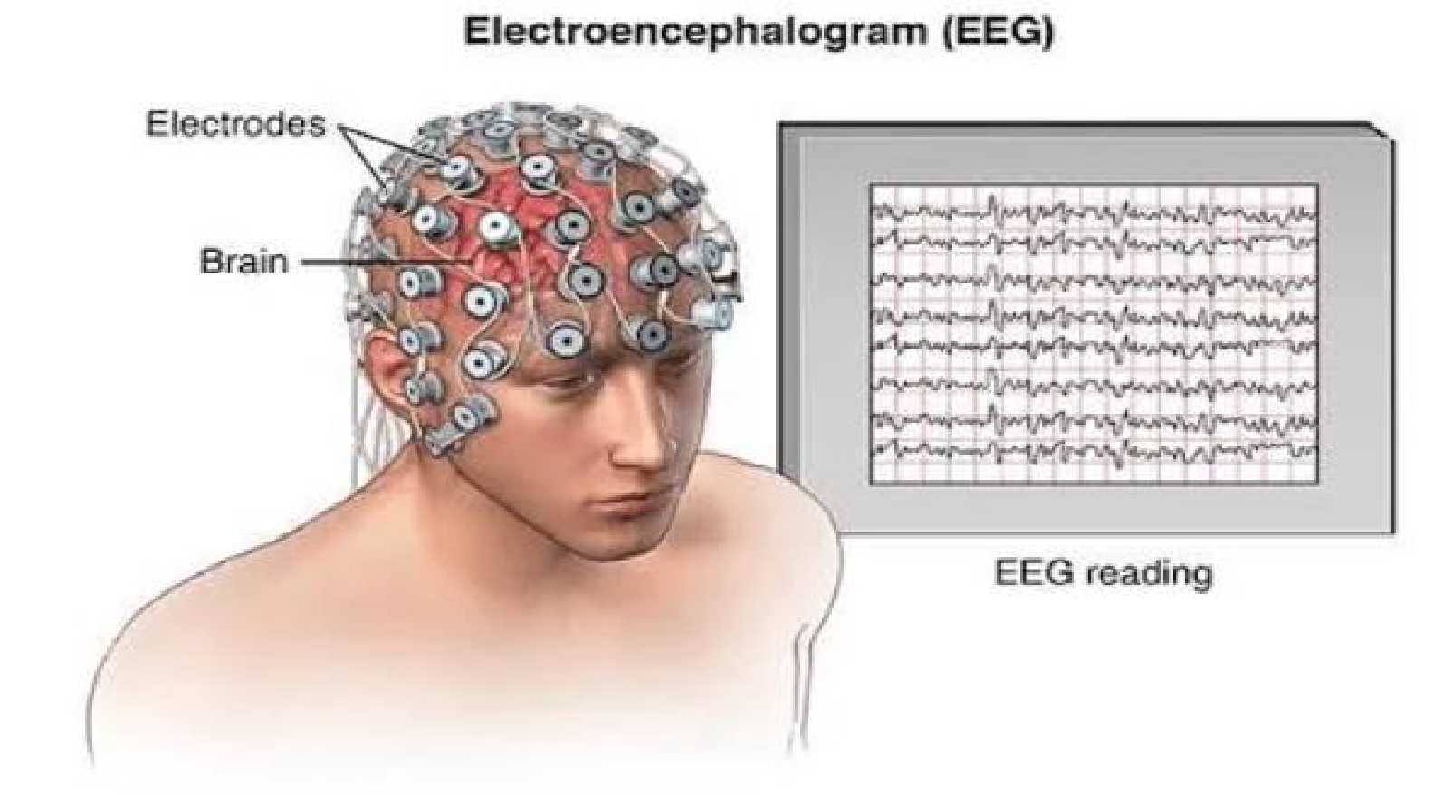
Emotion recognition based on electroencephalogram has been a lucrative task nowadays. Different procedures are executed to improve the computational power of emotion recognition frameworks utilizing electroencephalogram (EEG). There are several computational strategies that have performed incredibly well in emotion recognition task. An automated recognition system is typically limited to few emotions. So more effective and accurate emotion classification is the primary objective for Artificial Intelligence researchers. This paper aims to classify three emotion recognition conditions, such as positive, negative, and neutral. To reduce the over-fitting dispute, principal component analysis (PCA) is applied to extricate the most significant parts of input features. Besides, covariate move adjustment of the essential parts is executed to limit the non-stationary impact of EEG signals. Several machine learning models have been deployed to classify this problem. In this case, traditional algorithms such as logistic regression, linear support vector machine, random forest, artificial neural network, long short term memory (LSTM) and extreme gradient boosted (XGBoost) classifier have been enforced on the EEG brainwave signal dataset. The XGBoost has performed outstandingly in terms of accuracy and less time complexity.